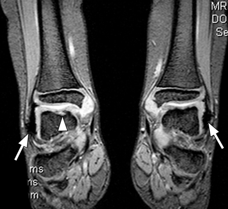
Figure 7a.
Hemophilic arthropathy. Coronal T2*-weighted fast GRE MR images (T2 FFE; Philips) (695/14; flip angle, 25°; bandwidth, 108.6 Hz/pixel; voxel size, 0.58 × 0.73 × 4.0 mm; FOV, 178 × 170 mm) of middle (a) and posterior (b) parts of ankle joints in a hemophiliac patient show dark areas of hemosiderin deposition (arrows in a and b) in both joints. The right talus bone shows irregularity and osteochondral change (arrowhead in a). T2*-weighted GRE MR images are useful in evaluation of hemophilic arthropathy because they show hemosiderin deposition, articular cartilages, and osteochondral changes well.

