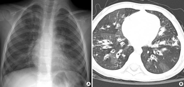
Fig. 1.
Chest radiographs on admission. (A) Chest plain radiograph shows diffuse reticulonodular densities in both central lung areas symmetrically. (B) On computed tomographic image with lung window setting, diffuse bronchiectasis is seen in both lungs. There are hyperlucent areas in the lung parenchyma due to peripheral bronchial obstruction.

