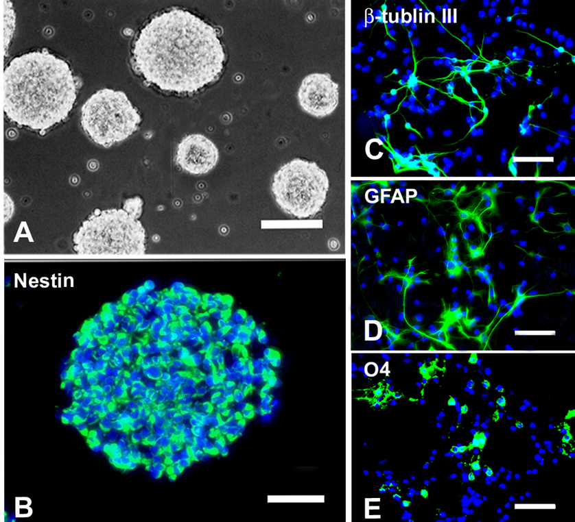
Fig. 2.

Morphological and immunocytochemical characteristics of NSCs. (A) Phase contrast photomicrograph of neurospheres cultured in growth medium supplemented with 20ng/ml EGF and 20ng/ml bFGF. (B) Cells in a neurosphere were immunopositive for nestin (green). (C~E) When cultured in medium containing no growth factors but 1% FBS for 5 days, NSCs differentiated into neurons (βIII-Tubulin+, C), astrocytes (GFAP+, D) and oligodendrocytes (O4+, E). Cells in B~E were counterstained with Hoechst 33342 (blue), a nuclear dye. A: bar = 200µm; B: bar = 50µm; C~E: bar = 25µm. (Adapted from Hu et al., J Neurosci Res 78:637–646, 2004)
