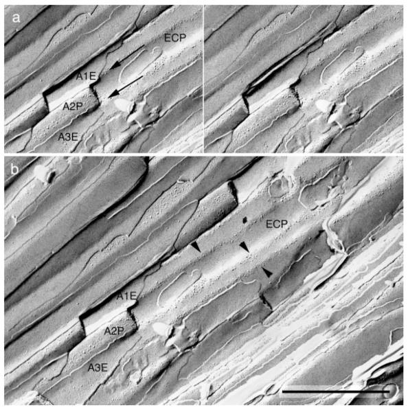
Fig. 4.
A stereomicrograph of a freeze-fracture replica from the edge of an olfactory nerve fascicle. a: A portion of the P face of an ensheathing cell surface (ECP) is indented by several overlying axons (A1–A3) that run parallel to each other. The fracture plane passed through the E face of axon 1 (A1E), the P face of axon 2 (A2P), and the E face of axon 3 (A3E). Ridges of the ensheathing cell (arrows) face extracellular space that lies between these adjacent axons. b: A more extensive view of the same field shows that intramembrane particles of the ensheathing cell are concentrated into linear arrays (arrowheads) along these ridges, while membrane area between the ridges is nearly devoid of IMP. Scale bar = 1 μm.

