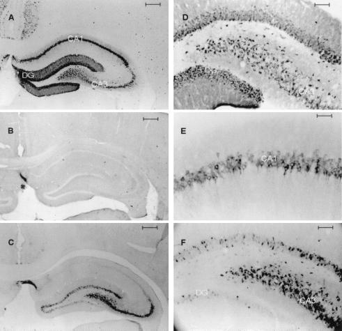
Figure 3.
Photomicrographs of COX2 immunocytochemistry from coronal sections through anterior hippocampus: 24 h after 15 min of global ischemia (A); 24 h after global ischemia, antibody preabsorbed with recombinant murine COX2 protein (B); 24 h after sham operation (C); hylar region, 24 h after global ischemia (D); CA1 24 h after global ischemia (E); and hylar region, 24 h after sham operation (F). [Bars = 400 μm (A–C), 50 μm (D and F), and 25 μm (E).]

