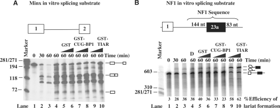
Figure 6.
CELF proteins block the splicing of NF1 exon 23a. (A) In vitro splicing assays were carried out using an in vitro-transcribed Minx RNA containing exons 1 and 2 and an intron in between (represented in the diagram) and HeLa nuclear extract. A splicing time course is shown in lanes 1–3. CELF proteins were added in increasing amounts. (B) In vitro splicing assays were carried out using an in vitro-transcribed RNA containing NF1 exon 23a and both upstream and downstream intronic sequence (represented in the diagram) and HeLa nuclear extract. A splicing time course is shown in lanes 1–3. The ability of CELF proteins to block the splicing of NF1 exon 23a was tested using increasing amounts of GST (0.75–1.5 µg), GST-CUG-BP1 (0.75–1.5 µg), or GST-TIAR (0.75–1.5 µg) and the splicing reaction was carried out for 60 min. Roeder D (labeled as D) with no protein was used as a negative control. The percentage of efficiency of lariat formation is given for the NF1 splicing assay.

