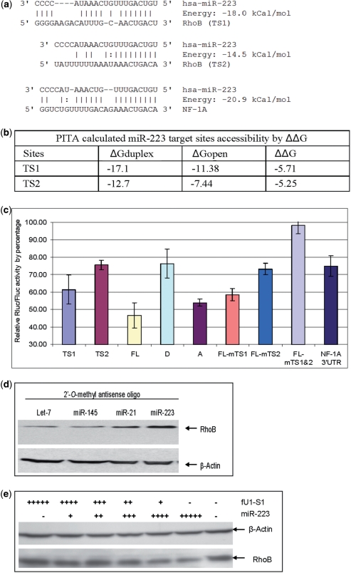
Figure 2.
RhoB is a miR-223 target and the two target sites contribute differentially to the total repression of RhoB translation. (a) miRnada predicted MiR-223 target sites in the 3′UTR of RhoB and NF-1A. (b) PITA calculated miR-223 target accessibility as measured by ΔΔG. TS1 has a slightly better accessibility than TS2. (c) Reporter assay of miR-223 inhibition of reporters carrying sequence fragments with miR-223 MREs. Rluc was fused with different fragments of the RhoB or NF-1A 3′UTR. The Y-axis represents relative expression of Rluc to Fluc when co-transfected with fU1-miR-223 and normalized to co-transfection with fU1-miR. Lane 1: MiR-223 target site one (TS1) short target sequence only; Lane 2: MiR-223 target site two (TS2) short target sequence only; Lane 3: FL RhoB 3′UTR (FL); Lane 4: First half of RhoB 3′UTR (D with TS1 only); Lane 5: Second half of RhoB 3′UTR (A) with TS2 only; Lane 6: FL with mutated TS1; Lane 7: FL with mutated TS2; Lane 8: FL with both TS1 and TS2 sites mutated; Lane 9: NF-1A 3′UTR. Each bar represents the average of at least three independent transfections with duplicate determinations for each construct. Error bars represent the standard deviation (SD). (d) Western blot result of RhoB expression in CEM cells when miR-223 function was blocked. CEM cells were transfected with a 2′-O-methyl anti-let-7 (lane 1), anti-miR-145 (lane 2), anti-miR-21 (lane 3), and anti-miR-223 (lane 4) oligo. Total cell extract were prepared 48 hours post transfection. The data show both anti-miR-223 and anti-miR-21 resulted in elevated RhoB protein levels while neither anti-let-7 nor anti-miR-145 antagomirs affected the RhoB protein level. Both miR-223 and miR-21 were predicted to target RhoB by miRanda and TargetScan (Figure 1b, miR-223 TS2 is located near miR-21 TS). (e) Western blot result of RhoB expression in Hela cells in the presence of miR-223. Hela cells were transfected with the miR-223 expression cassette. A U1 promoter-driven shRNA S1 (targeting the HIV Tat/Rev exon) was used as control. ‘−’ represents the absence of miR-223/S1 in the transfection. ‘+’ represents the present of miR-223/S1. The number of ‘+’s represents the amount of miR-223/S1 in the transfection. Total cell extracts were prepared 48 hours post transfection.

