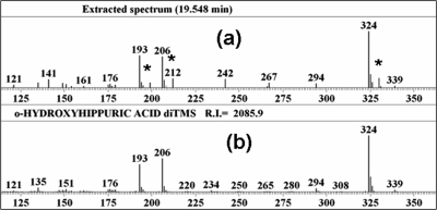
Figure 1.
Salicyluric acid in pooled urine sample: (a) partial mass spectrum obtained by gas chromatogrphaphy−mass spectrometry of a trimethylsilylated extract of pooled urine (isotopically labeled peaks are indicated by asterisks at m/z values 199, 212, and 330); (b) partial library mass spectrum of the di-trimethylsilyl derivative of standard salicyluric acid (o-hydroxyhippuric acid). Data were analyzed using the Automated Mass Spectral Deconvolution and Identification System (AMDIS; NIST, Gaithersburg, MD; http://chemdata.nist.gov/mass-sps/amdis).

