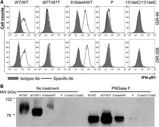
Figure 3.
Faint IFN-γR1 expression in P cells. (A) FACS analysis of IFN-γR1 on EBV-B cells from a healthy control (WT/WT), a patient with partial recessive IFN-γR1 deficiency (I87T/I87T), a patient with partial dominant IFN-γR1 deficiency (818del4/WT), patient P and a patient with complete recessive IFN-γR1 deficiency with no cell surface expression (131delC/131delC). Gray area: Isotypic control antibody; bold dark line: specific extracellular IFN-γR1 antibody (GIR-94 or GIR208). (B) Immunoprecipitation of IFN-γR1 from WT/WT, I87T/I87T, 818del4/WT, P and 131delC/131delC EBV-B cells, using a specific antibody recognizing the intracellular part of IFN-γR1 (C-20), with or without subsequent PNGase F treatment. The same antibody was used to detect IFN-γR1. Left panel: without PNGase F treatment; right panel: with PNGase F treatment.

