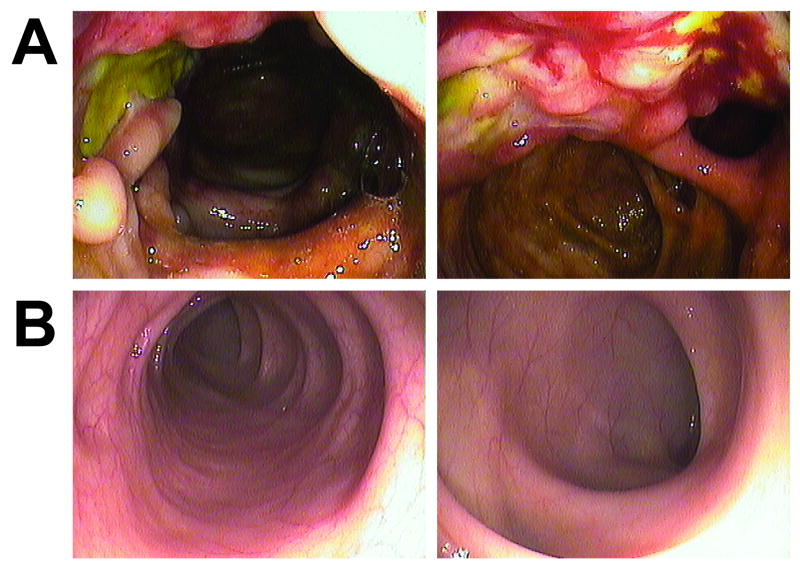
Figure 3.
Colonoscopy images from NEMO enterocolitis. A) Representative images from a study at Patient 3's initial presentation with abdominal symptoms, showing exudate (left frame) and mucosal inflammation (right frame). B) Two months after initiation of anti-inflammatory therapy, images were obtained from the same segment of bowel.

