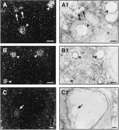
Figure 4.
Detection of DBH mRNA in monkey oocytes by in situ hybridization using a 35S-UTP DBH cRNA transcribed from the ovarian DBH cDNA shown in Fig. 1. (A) Oocytes from primary follicles (arrows) and preantral follicles (arrowheads). (B) Oocytes from three different sizes of preantral follicles (arrowheads). (C) Oocyte (arrow) of an antral follicle. (A1–C1) Bright-field photomicrographs of the dark-field images shown in A–C. The silver grains are not apparent in the bright-field pictures because they are out of the focus plane for the underlying tissue. (Bars = 75 μm.)

