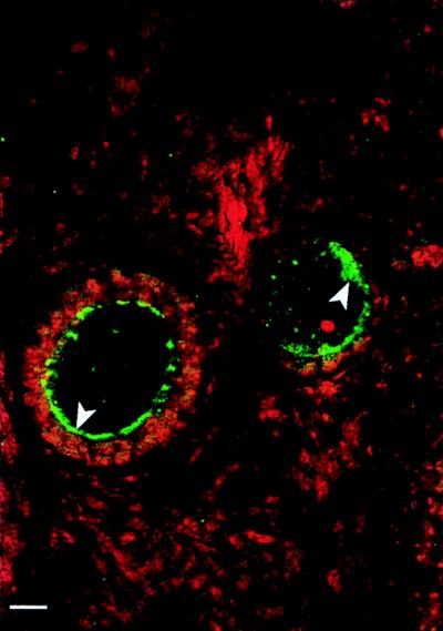
Figure 5.
Detection of DBH protein by immunohistochemistry/confocal microscopy in monkey oocytes. The DBH immunoreactivity (developed with fluorescein isothiocyanate to a green color) appears to be predominantly in subcellular granule-like structures adjacent to the cell membrane (arrowheads). Some immunoreactivity also is observed in the cytoplasm. The cell nuclei were stained (red) with propidium iodide, which binds to double stranded DNA. (Bar = 10.5 μm.)

