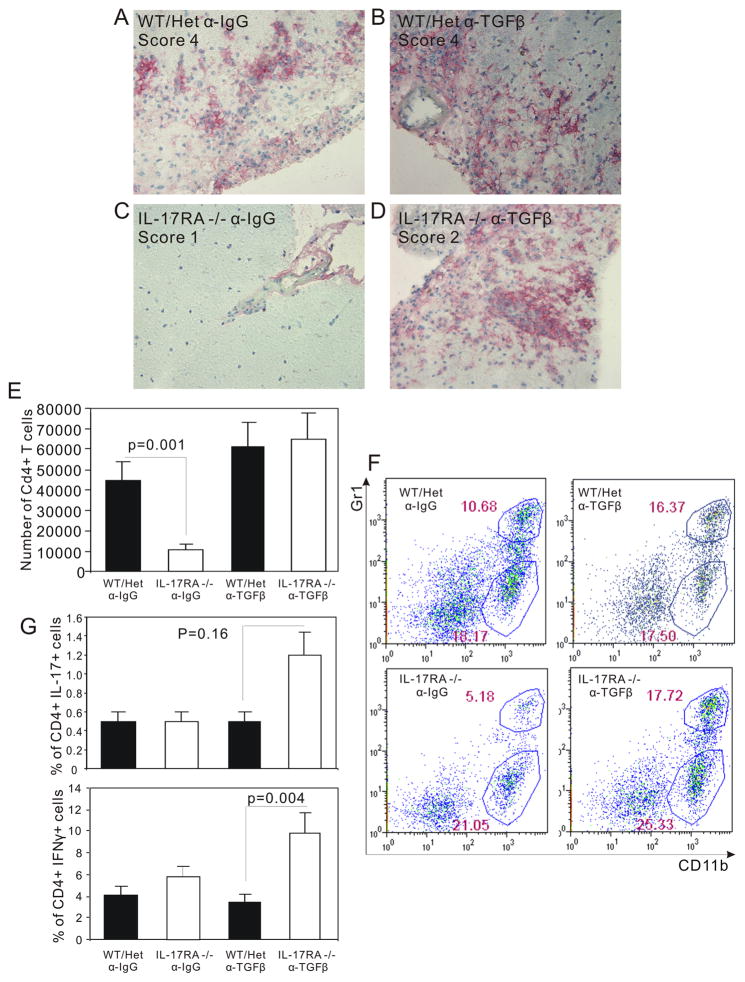
Figure 6.
Severe CNS inflammation in IL-17RA −/− mice treated with anti-TGFβ antibodies. Wild type mice (WT), IL-17RA +/− (Het), and IL-17RA −/− littermates were immunized with MOG35–55 peptide to elicit EAE. Anti-TGFβ antibodies were administered at day 5 and day 9. At 24 days post-immunization, immuno-histochemistry was done as described in Materials and Methods to determine the degree of leukocyte infiltration using anti-CD11b antibody. (A) IL-17RA +/− injected with control Ig, peak clinical score 4. (B) IL-17RA +/− injected with anti-TGFβ, peak clinical score 4. (C) IL-17RA −/− injected with control Ig, peak clinical score 1. (D) IL-17RA −/− injected with anti-TGFβ, peak clinical score 2. Wild type mice (WT), IL-17RA +/− (Het), and IL-17RA −/− littermates were immunized with MOG35–55 peptide to elicit EAE. Anti-TGFβ antibodies were administered at day 5 and day 9. Mice developed EAE were used around day 20 after the induction of EAE. The number of CD4+ T cells infiltrating CNS was calculated based on flow cytometry and cell count (E). CNS infiltrating cells were analyzed for Gr-1 and CD11b positive cells (F). CNS infiltrating lymphocytes were stimulated for 16 hours with MOG35–55 peptide, and then cells making IFNγ or IL-17 were analyzed by flow cytometry (G). The result was the average of data obtained from 4 independent experiments.

