
FIG. 2.
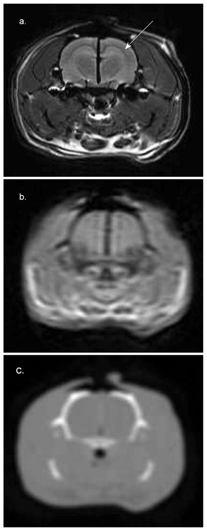
Cross section of the brain of a 19-week-old 546-g Sprague-Dawley rat. Panel a: 7T MR (FOV = 50 mm2). Panel b: 3T MR (FOV = 250 mm2). Panel c: CT (FOV = 320 mm2). The 7T image was acquired with an FOV large enough to include the surrogate fiducial system. The CT and 3T MR images were acquired with FOVs large enough to include the localizer box. These images were interpolated to a size comparable to the 7T image for comparison. The hippocampus is clearly visible in panel a (marked by the arrow) but cannot be identified in the other images. The dark line running in the vertical direction at midbrain in panels a and b is a piece of radiochromic film that was placed in the sagittal plane to verify the 7T Gamma Knife irradiation procedure.