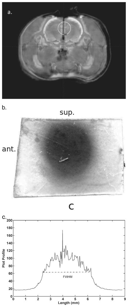
FIG. 6.
Panel a: Fusion of the 7T MR and CT images that was used to target the shot on the film in the cadaver rat brain. The centers of volume of specific features, such as bone and brain, agree to <2 voxels in all places. Panel b: The exposed film, which had been oriented in the animal such that the left side of the film (in the figure) was the anterior edge inside the animal and the top of the film was the superior edge. The LGP plan in panel a shows that the center of the shot was placed 4.5 mm from the inferior edge of the film. The measured distance from the inferior edge of the exposed film to the center of the shot, as indicated by the FWHM, was 4.5 mm. This shows that the shot was localized in the superior-inferior direction to <0.1 mm by the 7T LGP plan. Panel c: A profile through the horizontal axis of the exposed film. The FWHM was 5.1 mm, which shows that the shot was well centered along the X axis.

