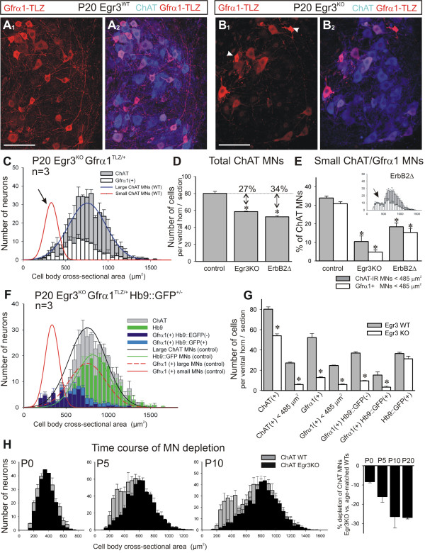
Figure 5.
Small diameter Gfrα1+ motor neurons are selectively lost in the muscle spindle mutant Egr3KO and ErbB2FLOX/-myf5Cre/+ animals. (A,B) Lamina IX confocal images from a P20 Egr3 wild type (A) and Egr3KO mutant (B). (A1, B1) Gfrα1-TLZ (red) and (A2,B2) superimposed with ChAT (blue). Small MNs intensely labeled with Gfrα1-TLZ are frequent in wild type but mostly absent in Egr3KO mutants. Somatic shrinkage is apparent in the few small Gfrα1-TLZ MNs found in Egr3KO mutants (arrowheads in B1). (C) Average size distribution of ChAT+ (gray bars) and Gfrα1-TLZ (white bars) MNs in P20 Egr3KO animals (n = 3 animals; error bars indicate SEM). Superimposed curves represent the wild-type distributions of small and large MNs. ChAT+ and Gfrα1-TLZ+ populations in Egr3KO mutants are both unimodal corresponding with large MNs. Small MNs are mostly absent (arrow). (D) Number of ChAT+ neurons sampled per ventral horn in 70-μm thick sections of P20 animals. Egr3KO (n = 3 animals) and Erb2Δ animals (n = 5) show significant depletions compared to controls (asterisks indicate P < 0.001, one-way ANOVA; P < 0.01 post-hoc Tukey-tests). (E) Percentages of the total ChAT+ population represented by 'small' (<485 μm2) MNs labeled with ChAT (gray bars) or Gfrα1-TLZ (white bars) in Egr3KO and Erb2Δ mutants. Both animals show a large depletion of small MNs compared to control (asterisks indicate P < 0.001, one-way ANOVA; P < 0.01 when compared to control using post-hoc Tukey-tests). In ErbΔ2 animals the reduction is not as pronounced as in Egr3KO animals despite a larger depletion in total ChAT+ MNs (D). Inset shows the average size distribution histogram in ErbΔ2 animals (gray bars, ChAT+; white bars, Gfrα1+) suggesting partial depletion of both large and small MNs. (F) Average size distribution of MNs in Egr3KO animals with different combinations of markers: ChAT+ (gray bars), Hb9::GFP+ (green bars), Gfrα1-TLZ+/Hb9::GFP- (dark blue bars) and Gfrα1-TLZ+/Hb9::GFP+ (light blue bars). Superimposed are the fitted distributions for different types of MNs in the wild-type. (G) Average number of MNs per ventral horn in each category. Asterisks denote significant differences (P < 0.001, t-test) between wild-type (gray bars) and Egr3KO mutants (white bars). Significant differences were observed in all MNs and in small MNs (<485 μm2) labeled with either ChAT or Gfrα1-TLZ. Hb9::GFP+ MNs are not significantly depleted in Egr3KO mutants, though Gfrα1-TLZ expression is downregulated in surviving large diameter MNs (F). (H) Time course of size differentiation and depletion of small MNs in Egr3KO animals. Gray bars indicate size distribution in wild type (WT; n = 3 animals for each age) and black bars in age matched Egr3KO animals (n = 2 at P0, n = 4 at P5 and P10). At P0, MN sizes are unimodal and there is a small depletion in ChAT+ neurons distributed in all size bins. At P5 there is initial differentiation of small vs. large MNs and in the Egr3KO mutant there is a larger depletion of ChAT+ MNs concentrated in the small bins. At P10 the size distribution in the wild type resolves into two discrete peaks for the small and large population and in the Egr3KO mutant the depleted neurons are clearly in the small size bins. Histogram at the right show the percentage depletions calculated in Egr3KO mutants of different ages. Scale bars: (A1, B1) 100 μm.

