
Figure 1.
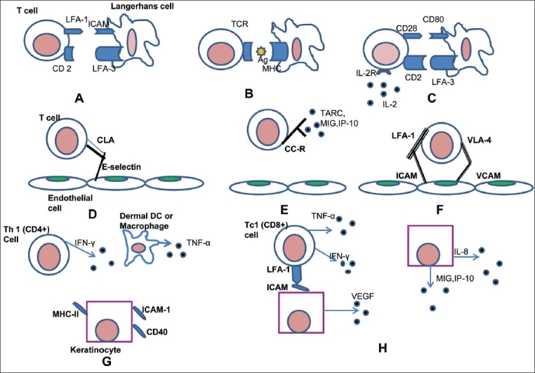
The current hypothesis by which T cells get activated and how the release of various mediators leads to the hyperproliferation of the keratinocyte. (Modified from Mehlis and Gordon1, Krueger37) (A) T cell binds to an Antigen-Presenting Cell, (B) T cell receptor recognizes the antigen presented on MHC of the APC in an antigen specific interaction, (C) Non-antigen specific cell-interaction. The stimulation of both TCR and CD28 pathways lead to transcription of IL-2, TNF-α, GM-CSF and IFN-Y, (D) T cell is rolling on the endothelium, (E) T cell surface proteins are activated, (F) T cell binds to the endothelium and diapedesis occurs, (G) Dermal Th1 cells release IFN-Y and other cytokines, which lead to increases expression of inflammatory and adhesion proteins on keratinocytes, (H) Keratinocytes proliferate; synthesize angiogenic cytokines / chemokines that cause leukocyte trafficking and increase leukocyte adhesion to the endothelial cells.