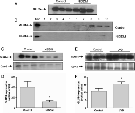
Figure 5.
Myocardial glucose transporter 4 (GLUT4) levels in patients with non-insulin-dependent diabetes mellitus or left-ventricular dysfunction. (A) Immunoblot of GLUT4 levels in total cell lysates from patients with non-insulin-dependent diabetes mellitus and controls. (B–D) Sucrose gradient fractionation of cardiac muscle proteins. (B) Upper panel, representative immunoblot of GLUT4 expression at the plasma membrane fraction (Mbn) and across sucrose gradient fractions (1–10) in a control patient. Lower panel, immunoblot of GLUT4 expression at the Mbn and across sucrose gradient fractions (1–10) in a patient with non-insulin-dependent diabetes mellitus. (C) Immunoblot of GLUT4 levels at the plasma membrane fraction (upper panel) and Caveolin-3 (Cav-3) as loading control (lower panel) in controls or patients with non-insulin-dependent diabetes mellitus. (D) Quantification of GLUT4 expression at the sarcolemma in control patients (n = 3) and patients with non-insulin-dependent diabetes mellitus (n = 3) in arbitrary optical units; (*P = 0.01). (E and F) Myocardial GLUT4 levels in patients with left-ventricular dysfunction. (E) Immunoblot of GLUT4 levels at the sarcolemma (upper panel) and Caveolin-3 as loading control (lower panel) in control patients or patients with left-ventricular dysfunction. (F) Quantification of GLUT4 expression at the sarcolemma in control patients (n = 3) and patients with left-ventricular dysfunction (n = 4) in arbitrary optical units; *P = 0.01.

