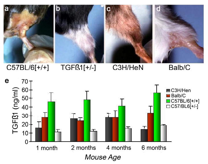
Figure 3.

TGFβ1 is among the most studied circulating biomarkers of radiation fibrosis, and there is a strong association between circulating levels of this cytokine and fibrosis in irradiated mice and in human disease. Panels a-d show mice irradiated to the hind limb with 40 Gy at 4 weeks post-exposure. Note the depilation of the C57BL/6[+/+] and Balb/C mice compared to the relatively healthy appearance of the C3H/HeN and heterozygous knockout TGFB1[+/-] mice. The C57Bl/6 mouse is known to have a high propensity for radiation fibrosis and the C3H/HeN a low, while the Balb/C is intermediately fibrosing following irradiation. Heterozygous knockout TGFβ1 mice are phenotypically normal, but have a life-long low level of circulating TGFβ1 and a similarly low level of depilation, muscle wasting, and scarring following irradiation. The histogram (panel e) indicates the naturally occurring plasma levels of TGFβ1 (no radiation given) for these 4 mouse strains as a function of age.
