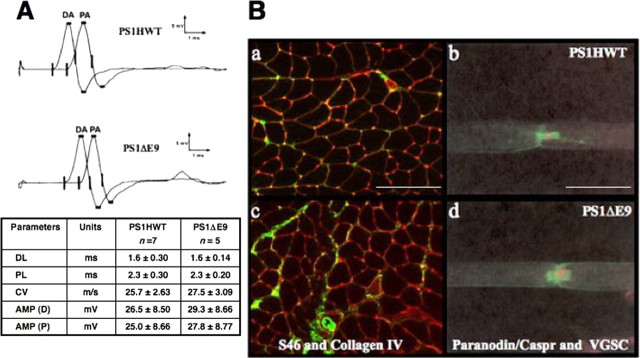
Figure 4.
Conduction velocity, muscle tissue morphology, and myelination are intact in sciatic nerve of FAD-linked PS1 variants. A, CMAPs were recorded after distal and proximal stimulation of sciatic nerves of transgenic mice harboring FAD-linked PS1HWT and PS1ΔE9. Waveform, latencies, and amplitudes of CMAPs were found to be similar in both groups. DL, Distal latency; PL, proximal latency; CV, conduction velocity; AMP (D), distal amplitude (DA); AMP (P), proximal amplitude (PA). Ba, Bc, Muscle sections of PS1HWT (a) and FAD-linked PS1ΔE9 (c) mice. Immunostaining with antibodies raised against slow-tonic myosin heavy chain isoforms, indicating muscle morphology (green), and with antibodies raised against collagen IV (red) revealed no signs of muscular atrophy. Scale bar, 10 μm. Bb, Bd, Nodal and paranodal structures of sciatic nerves of PS1HWT (b) and FAD-linked PS1ΔE9 (d) mice were visualized using antibodies raised against pan VGSC (red) and antibodies raised against paranodin/Caspr (green), respectively. No morphological abnormalities could be detected in PS1ΔE9 mice. Scale bar, 50 μm.

