Figure 5.
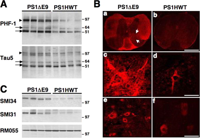
Neuropathology in lumbar spinal cord of FAD-linked PS1ΔE9 mice. A, Western blot analysis of tau phosphorylation in detergent-soluble protein extracts of lumbar spinal cord of PS1HWT and PS1ΔE9 mice. Note the increased immunoreactivity for PHF-1 in the spinal cord of PS1ΔE9 compared with PS1HWT mice. No significant change in the expression of total tau (Tau5) was observed between PS1ΔE9 and PS1HWT mice samples. Tau isoforms of medium (arrows) and high (arrows) molecular weight are indicated. B, Distribution and localization of PHF-1 expression in lumbar spinal cord sections of PS1ΔE9 and PS1HWT mice. Low-power (a, b) and high-power (c–f) images of PHF-1 immunoreactivity in spinal cord sections of PS1HWT (b, d, f) and PS1ΔE9 (a, c, e) show increased PHF-1 immunoreactivity in spinal cord sections of PS1ΔE9 mice. Higher levels of PHF-1 immunoreactivity were detected in white matter and in cell bodies in the ventral horn (arrowheads). Scale bars: a, b, 750 μm; c, d, 110 μm; e, f, 65 μm. C, Phosphorylation of NF-M is markedly increased in detergent-soluble protein extracts of lumbar spinal cord of PS1ΔE9 mice compared with PS1HWT mice, as shown by SMI34 and SMI31immunoreactivity. These antibodies recognize a phosphorylated epitope on both NF-H and NF-M subunits. Note that not all phospho-epitopes on NF-M are altered in PS1ΔE9, as shown by RM055, a monoclonal phospho-specific antibody that binds to a phosphorylated epitope in the tail domain of NF-M (Virginia Lee, personal communication).

