Figure 1.
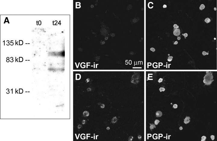
Upregulation of VGF immunoreactivity in cultured DRG neurons. A, In Western blot analysis, samples from t24 DRG cultures show a band at ∼90 kDa, which corresponds to the unprocessed form of VGF. The lower-molecular-weight bands most likely correspond to proteolytic fragments of VGF. Consistent with VGF increase at t24 compared with t0, VGF-immunoreactive bands at t0 are nearly below the detection limit. B, C, Cultured DRG neurons fixed at t0 and double labeled with anti-VGF (B) and anti-PGP (C), which shows all neurons in the field of view. D, E, Cultured DRG neurons fixed at t24 and double labeled with anti-VGF (D) and anti-PGP (E). The number of VGF-positive neurons increased at t24 (D).

