
Figure 4.
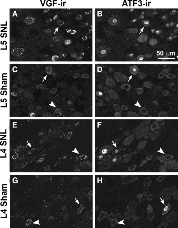
Relationship of VGF- and ATF3-IR in L5 and L4 DRG 1 d after SNL or sham surgery. In double-labeled sections, VGF- and ATF3-IR overlapped substantially in injured L5 DRG (A, B). In sham L5 DRG (C, D), in L4 DRG from SNL animals (E, F), and in sham L4 DRG (G, H), VGF-IR was present in ATF3-positive and ATF3-negative neurons. Arrows show examples of colocalization of VGF-IR and ATF3-IR. Arrowheads show examples of VGF-positive neurons without ATF3-IR in their nuclei.