Figure 5.
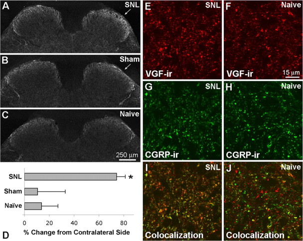
Changes in VGF-IR in L5 segment of spinal cord after SNL. A–C, Three days after SNL, VGF-IR appeared increased within the dorsal horn ipsilateral to SNL (A) compared with sham surgery (B) and naive animals (C). D, Quantitative analysis indicated a significant increase in VGF-IR ipsilateral to SNL compared with sham surgery and naive spinal cord (one-way ANOVA and Student–Newman–Keuls multiple comparisons test, p < 0.05; n = 4 for each group). E–J, Compared with naive spinal cord (F, H, J), VGF-IR (red) was increased after SNL (E, G, I), whereas CGRP-IR (green) appeared unchanged at this time point. The colocalization of VGF- and CGRP-IR in superficial dorsal horn was also increased after SNL as indicated by the presence of more yellow and orange puncta ipsilateral to SNL (I) compared with naive spinal cord (J).

