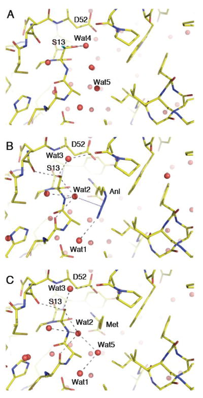
Figure 3.
Binding of azidonorleucine and methionine to MetRS-SLL. The three panels show stick representations of the amino acid binding sites of free or liganded MetRS-SLL, all in the same orientation. Carbon atoms are in yellow, nitrogen atoms in blue and oxygen atoms in red. Water molecules are drawn as red spheres. Relevant hydrogen bonds are indicated by grey dotted lines. Panel A: unliganded MetRS-SLL; the two conformations of the side chain of S13 are drawn, with conformation 1 in standard colours and conformation 2 with the hydroxyl oxygen in green. Panel B: MetRS-SLL:Anl complex. Only the major conformation of S13 is represented. The electrostatic interaction between Wat2 and the N2 atom of Anl is symbolized by a blue dotted line. Panel C: MetRS-SLL:Met complex. Only the major conformation of S13 is represented.

