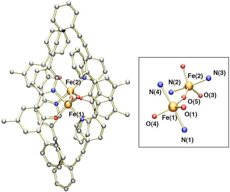
Figure 7.

A ball and stick representation of the low-resolution X-ray crystal structure of [Fe2(μ-O)(LMe,Ph)2] (2). The coordination environment of the μ-oxodiiron(III) core is shown on the right. Due to the poor quality of the X-ray data, only the atom connectivity of the structure could be obtained. The atoms are color coded according to the following: gray = carbon, red = oxygen, blue = nitrogen, and orange = iron.
