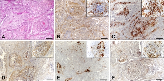
Fig. 1.
Immunohistochemical markers in canine seminoma. (A) Seminomas consisted of aggregates of germ cells that filled the affected seminiferous tubules. The tumor cells were large and polyhedral with vesicular nuclei and prominent nucleoli. H&E stain. (B-F) Positive signals to tumor cells; (B) alpha-fetoprotein, (C) inhibin-alpha, (D) vimentin, (E) desmin and (F) c-KIT. Immunostain and counterstain with Harris hematoxylin. Scale bars = A: 350 µm, B-F: 140 µm.

