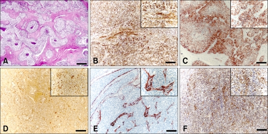
Fig. 2.
Immunohistochemical markers in canine Sertoli cell tumors. (A) Cells within the tumor resemble Sertoli cells that normally populate the seminiferous tubules and are arranged into sheets or tubules separated by fibrous connective tissues. H&E stain. (B-F) Positive signals to tumor cells; (B) alpha-fetoprotein, (C) inhibin-alpha, (D) vimentin, (E) desmin and (F) c-KIT. Immunostain and counterstain with Harris hematoxylin. Scale bars = A: 350 µm, B-F: 140 µm.

