Fig. 1.

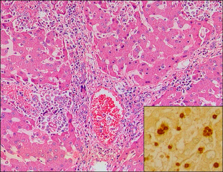
Liver of aborted sow, showing multifocal necrotic hepatitis. H&E, ×200. Insert: Note brown tachyzoites of Toxoplasma gondii in sinusoid and Kupffer cell. ABC stain, ×400.

Liver of aborted sow, showing multifocal necrotic hepatitis. H&E, ×200. Insert: Note brown tachyzoites of Toxoplasma gondii in sinusoid and Kupffer cell. ABC stain, ×400.