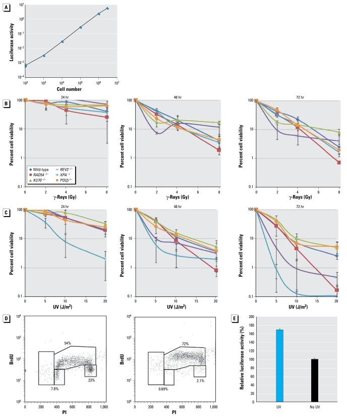
Figure 2.
Analysis of cell number by measuring ATP in cellular lysates. (A) The relationship in the range from 102/mL to 106/mL DT40 cells with luciferase activity (the amount of ATP). (B, C) Luciferase activity 24, 48, and 72 hr after irradiation with γ-rays (B) and UV (C); the pattern of cellular proliferation was very similar between 48 and 72 hr after γ-irradiation (B). (D) The cell cycle 6 hr after exposure to 5 J/m2 UV (right) and with no UV irradiation (left) by BrdU pulse-labeling. (E) Amount of luciferase activity per cell. UV irradiation augmented the amount of ATP by 70% at 24 hr after UV compared with nonirradiated cells; 104 cells were exposed to 5 J/m2 UV and subsequently incubated in 5 mL medium for 24 hr.

