Figure 1.
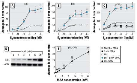
Activation of the CMV promoter by MAA in vitro. (A) HeLa cells were transfected with a human ERβ expression vector, the estrogen-responsive 3X-ERE-TATA-Luc reporter plasmid, and the CMV-β-gal reporter plasmid, treated for 18 hr with vehicle, increasing concentrations of E2, 5 mM MAA, or increasing concentrations of E2 plus 5 mM MAA, and assayed for luciferase activity. Data represent the average fold over control (± SE) of duplicate samples from three independent experiments. (B) HeLa cells were transfected with a human ERα expression vector, 3X-ERE-TATA-Luc, and CMV-β-gal and treated identically to the cells in (A). Data represent the average fold over control (± SE) of duplicate samples from three independent experiments. (C) MCF-7 cells were transfected with the 3X-ERE-TATA-Luc reporter plasmid and the CMV-β-gal reporter plasmid with and without the human ERα expression vector. The cells were treated as described for (A). Data represent the average fold over control (± SE) of duplicate samples from three independent experiments. (D) HeLa cells were transfected with a human ERα expression vector and treated with either vehicle or increasing concentrations of MAA for 18 hr. ERα protein expression was analyzed by Western blot. Data are representative of three independent experiments. (E) HeLa cells were transfected with the pRL-CMV reporter plasmid, treated with either vehicle or increasing concentrations of MAA for 18 hr, and assayed for luciferase activity. Data represent the average fold over control (± SE) of duplicate samples from three independent experiments.
*p < 0.05 compared with E2 treatment. **p < 0.05, and #p < 0.01, compared with E2 treatment. ##p < 0.01 compared with vehicle control.

