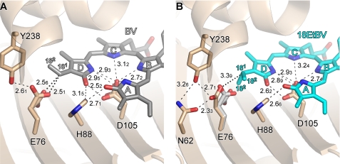
FIGURE 4.
Close-up views around the tetrapyrrole d-rings. A, PcyA-BV complex. B, PcyA-18EtBV complex. Stick models are shown for the substrate and the side chains nearby the D-rings. In B, Glu-76 of PcyA-BV is superimposed in gray on PcyA-18EtBV to clarify the conformational change upon reduction. The side chain of Asn-62 is not shown in A because it does not participate in hydrogen bonding with Glu-76 in the PcyA-BV complex. The Glu-76 side chain in PcyA-BV (A) and the ethyl group of the 18EtBV D-ring in PcyA-18EtBV (B) each adopt two conformations. Hydrogen bonds are drawn with dashed lines, whereas notable interactions are shown with dotted lines. Numerals represent the interatomic distances in Å unit. Due to limited accuracy of the interatomic distances the second decimal numerals are shown in subscript.

