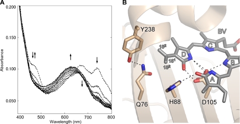
FIGURE 5.
Biochemical reaction of E76Q mutant protein and the close-up view of BV in E76Q-BV complex. A, analysis of BV reduction by E76Q mutant protein from Synechocystis sp. PCC6803 PcyA. Absorption spectra were monitored for 10 min with a 1-min interval. The initial spectrum is shown by a dashed line. The double arrow indicates the increase and decrease of the absorbance at 468 nm. The single arrows indicate the absorbance increase at 630 nm and indicate the absorbance decreases at 670 and 740 nm. B, close-up view around the BV D-ring in the E76Q-PcyA complex. BV, Gln-76, His-88, Asp-105, and Tyr-238 are shown by stick models. The vinyl group of the BV D-ring in E76Q-BV adopted two conformations. Hydrogen bonds are drawn with dashed lines. The interatomic distance between C181 and Gln-76 Oϵ is 3.47Å.

