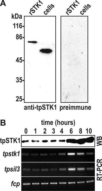
FIGURE 2.
Cell cycle-specific expression of native tpSTK1. A, Western blots probed with anti-tpSTK1 IgG (0.2 μg/ml) and preimmune IgG (0.2 μg/ml) that were isolated from the same rabbit. Lanes labeled rSTK1 contained 100 ng of purified recombinant tpSTK1; lanes labeled cells contained lysates from 1.3 × 10⁶ T. pseudonana cells. B, time course of tpSTK1 protein and tpstk1 mRNA expression in synchronized T. pseudonana cells. The times refer to hours after addition of silicic acid to silicon-starved cells. Formation of valve silica is at maximum between 4 and 6 h (39). For each time point, SDS-lysed cells from equal culture volumes were used for Western blot analysis (WB) and cDNA synthesis. The Western blot was probed with 0.2 μg/ml anti-tpSTK1 IgG. Reverse transcription (RT)-PCRs were performed from identical amounts of cDNA using primers specific for the indicated genes: tpsil3, gene encoding silaffin tpSil3; fcp, gene encoding a fucoxanthin chlorophyll-associated protein that is constitutively expressed in T. pseudonana under constant illumination (A. Scheffel and N. Kröger, unpublished communication).

