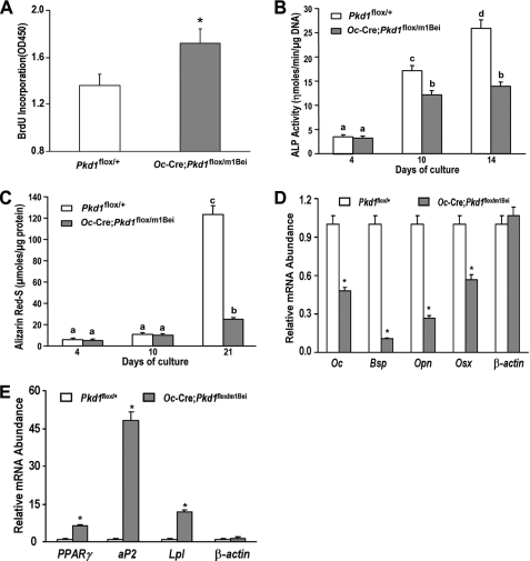
FIGURE 4.
Pkd1Oc-cKO osteoblasts have a developmental defect ex vivo. A, bromodeoxyuridine (BrdU) incorporation. Primary cultured Pkd1Oc-cKO osteoblasts exhibited a higher bromodeoxyuridine incorporation than control Pkd1flox/+ osteoblasts for 6 h, indicating increased proliferation in the Pkd1Oc-cKO osteoblasts. B, alkaline phosphatase (ALP) activity. Primary cultured Pkd1Oc-cKO osteoblasts displayed time-dependent increments in alkaline phosphatase activities for 14 days of culture, but the alkaline phosphatase activity was significantly lower at different time points compared with control Pkd1flox/+ osteoblasts. C, quantification of mineralization. Alizarin Red-S was extracted with 10% cetylpyridinium chloride and quantified as described under “Experimental Procedures.” Primary cultured Pkd1Oc-cKO osteoblasts had time-dependent increments in Alizarin Red-S accumulation for 21 days of culture, but the accumulation was significantly lower at different time points compared with control Pkd1flox/+ osteoblasts. D and E, gene expression profiles by real-time RT-PCR. 10-day cultured Pkd1Oc-cKO osteoblasts in osteogenic differentiation medium showed a significant attenuation in osteogenesis compared with control osteoblasts, as evidenced by a significant reduction in osteoblastic markers, including osteocalcin (Oc), bone sialoprotein (Bsp), osteopontin (Opn), and osterix (Osx). However, a marked increase of adipocyte markers, such as PPARγ, aP2, and Lpl, was observed from the Pkd1Oc-cKO osteoblasts under the same differentiation medium when compared with control osteoblasts. Data are mean ± S.D. from triple three independent experiments. *, significant difference from control (Pkd1flox/+) mice at p < 0.05. Values sharing the same superscript in B and C are not significantly different at p < 0.05.

