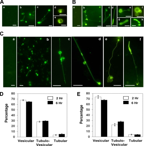
FIGURE 2.
GFP-LC3-positive tubulovesicular structures in cell lysates. A and B, HEK-293-GFP-LC3 (A) or HCT-116-GFP-LC3 (B) cells were treated with normal medium (panels a in A and B) or EBSS (rest of the panels) for 4 h. Cells were then lysed, and LC3-TVS were identified. Representative LC3-TVS are shown. Scale bar, 2 μm. C, HEK-293-GFP-LC3 cells treated with medium (panel a) or CPP for 3 h (panels b–f) were lysed and observed. Representative LC3-TVS are shown. Scale bar, 5 μm. D and E, HEK-293-GFP-LC3 cells were treated with CPP (D) or EBSS (E) for 2 or 6 h as indicated before being lysed. Three types of LC3-TVS were quantified in the lysates in randomly selected fields using fluorescence microscopy, and the percentages (mean ± S.D.) of each type of LC3-TVS were determined. Vesicular LC3-TVS are single ring-like structures (e.g. A, panels d and e); tubular LC3-TVS are tubules only (e.g. B, panel g); and tubulovesicular LC3-TVS are the combination of vesicular and tubular structures (e.g., A, panel c; B, panel b; B, panel d; B, panel e; and C, panels c–f).

