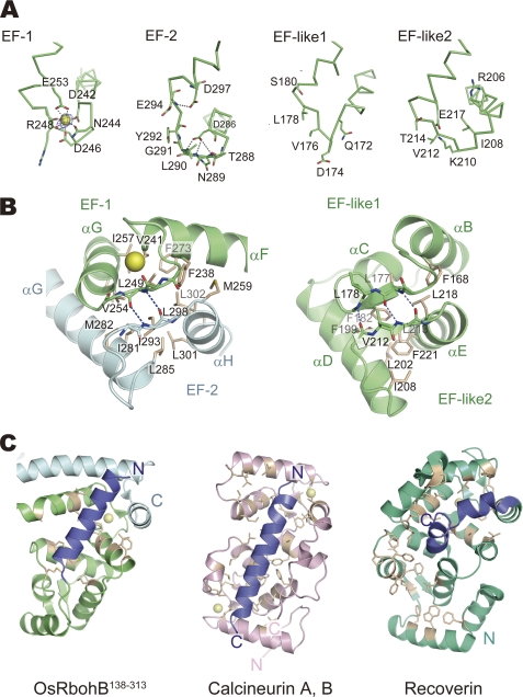
FIGURE 2.
EF-hand motifs of OsRbohB-(138–313) and similar proteins. A, representation of EF-hand motifs (EF-1, EF-2, EF-like 1, and EF-like 2). Difference Fourier maps showing contour levels higher than 5 σ. Ca2+ ion and water molecules are represented as a yellow and red spheres, respectively. Amino acid residues of EF-hand motifs at positions X to Z are shown as sticks. B, magnified view of EF-hand pairs composed of EF-1 and EF-2, and EF-like 1 and EF-like 2, respectively. Residues forming the hydrophobic cores are shown as white sticks. C, hydrophobic pockets of OsRbohB-(138–313), calcineurin B, and recoverin. Residues forming the hydrophobic pocket are shown as white sticks. The pocket of OsRbohB-(138–313) formed by swapped EF-hands and EF-hand-like motifs is occupied by an N-terminal helix (blue) protruding from the core domain. The pockets of calcineurin B and recoverin are occupied by α-helices protruding from calcineurin A and the C-terminal region (blue), respectively.

