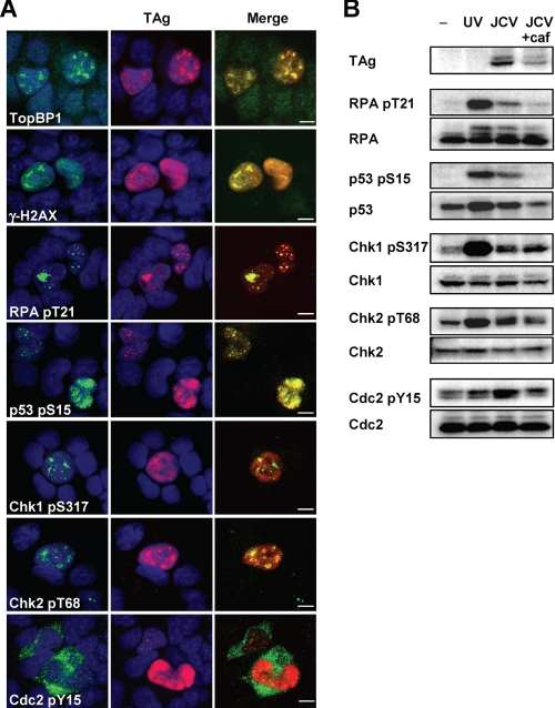
FIGURE 3.
Induction of G2 checkpoint signaling in JCV-infected IMR-32 cells. A, JCV-infected IMR-32 cells (14 d.p.i.) were subjected to double immunofluorescence staining with antibodies to the indicated phosphorylated forms of checkpoint proteins (green) and with antibodies to TAg (red). Cell nuclei were stained with DAPI (blue). TopBP1 indicates topoisomerase-binding protein-1; γ-H2A.X indicates phosphorylated histone H2A.X; RPA indicates RPA32. pS, phosphoserine; pY, phosphotyrosine; pT, phosphothreonine. Scale bars, 5 μm. B, JCV-infected IMR-32 cells (14 d.p.i.) incubated in the absence or presence of 2.5 mm caffeine (caf) for 15 h, as well as UV-treated or control (−) IMR-32 cells, were subjected to immunoblot analysis with antibodies to phosphorylated or total forms of the indicated proteins.

