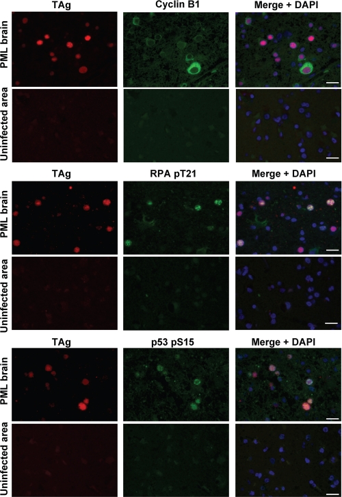
FIGURE 9.
TAg-expressing oligodendrocytes in the PML brain are arrested in G2 phase. Sections of brain tissue from an individual with PML were subjected to double immunofluorescence staining with antibodies to TAg (red) as well as with those to cyclin B1, Thr21-phosphorylated RPA32, or Ser15-phosphorylated p53 (green). Cell nuclei were counterstained with DAPI (blue). Images were obtained from JCV-infected and uninfected areas of brain tissue. Scale bars, 20 μm.

