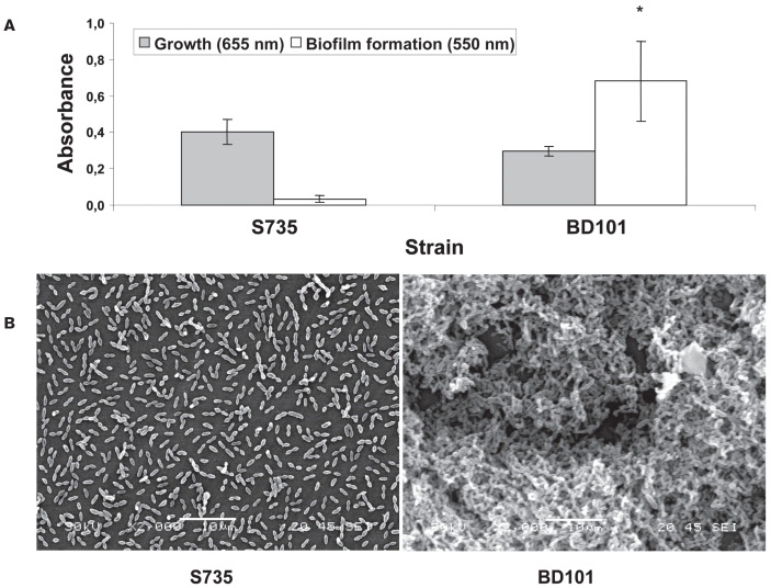
Figure 1.
Biofilm formation by Streptococcus suis S735 and its unencapsulated mutant BD101. (A) Crystal violet-stained biofilm following growth in polystyrene microplate. (B) Scanning electron micrographs of S. suis biofilms formed after 24 h of growth. * indicates a significant difference (P < 0.01) with the Student’s t-test between S. suis S735 and its unencapsulated mutant BD101.

