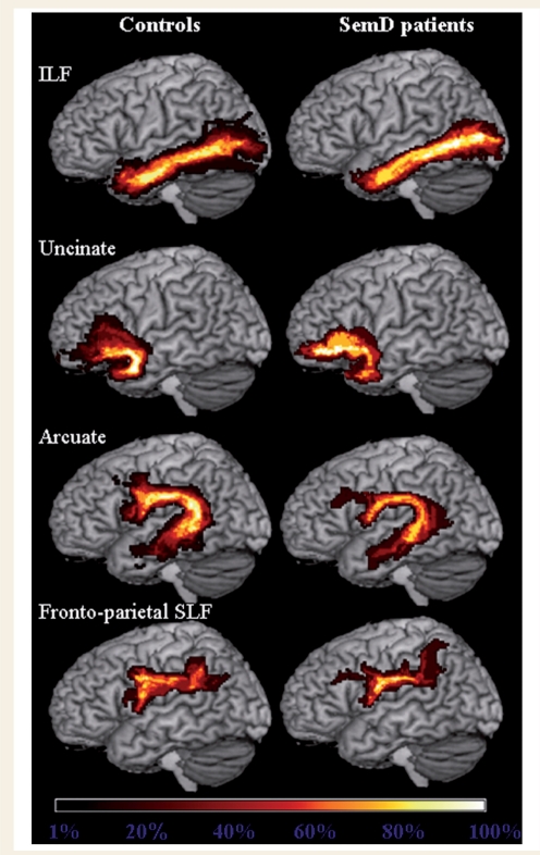
Figure 3.
Group probabilistic maps of the language-related white matter tracts from healthy controls and semantic dementia (SemD) patients. Left ILF, uncinate fasciculus, arcuate fasciculus and fronto-parietal SLF are superimposed on the three-dimensional rendering of the MNI standard brain. The colour scale indicates the degree of overlap among subjects.

