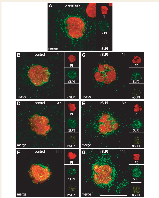
Figure 4.
SLPI localizes to the nucleus of circulating leukocytes isolated after recombinant mouse SLPI treatment. In all figures, ‘PI’ (red) shows the 3D reconstruction of the nucleus (labelled with propidium iodide), ‘SLPI’ (green) shows the total intracellular SLPI immunoreactivity, and ‘nSLPI’ (yellow) shows the SLPI immunoreactivity within the nucleus only. (A) A very small amount of SLPI is localized to the nucleus of leukocytes obtained from mice prior to contusion injury. (B, D and F) Vehicle-treated mice at 1, 3 and 11 h after injection show little change in nuclear SLPI. (C, E and G) In contrast, there is a marked increase in total intracellular and nuclear SLPI in leukocytes obtained from recombinant mouse SLPI-treated mice as early as 3 h and up to 11 h after injection. Vehicle and recombinant mouse SLPI were injected 1 h after spinal cord injury. All scale bars = 10 µm.

