Fig. 3.
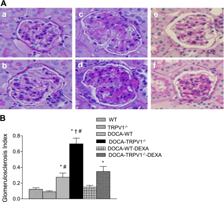
Representative light micrographs of kidney sections (A), stained with periodic acid-Schiff technique, from control WT and TRPV1−/− mice (a and b), and DOCA-salt-treated WT and TRPV1−/− mice with (e and f) or without (c and d) DEXA treatment. Bar graph (B) shows the average of glomerulosclerosis index. Values are means ± SE (n = 7 to 8). *P < 0.05 compared with control WT or TRPV1−/− mice. †P < 0.05 compared with DOCA-salt-treated WT mice. #P < 0.05 compared with DOCA-salt-treated WT or TRPV1−/− mice with DEXA treatment.

