Fig. 2.
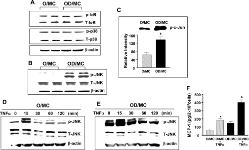
Elevated phosphorylated JNK levels and JNK activity in aging diabetic mesangial cells. Two separate isolates of mesangial cells from O and OD were placed in a serum-free condition for 24 h before protein collection. A: levels of phosphorylated IκB, total IκB, phosphorylated p38, total p38, and β-actin were measured by Western blots in a same membrane probed consecutively with respective antibody. Representative gels from 3 independent measurements of 2 separate cell lines show that the levels of phosphorylated IκB and p38 were similar between O/MC and OD/MC. B: levels of phosphorylated JNK were visibly increased in OD/MC. C: kinase assay demonstrated that increased phosphorylated JNK in OD/MC was associated with increased JNK activity. Representative gel of kinase assay of O/MC and OD/MC was shown on the top. The lower bars were the relative density of bands from the measurement of 2 mesangial cell lines from each group (n = 2). &P < 0.05 vs. O/MC. D and E: serum-starved old control and diabetic mesangial cells were treated with TNF-α (10 ng/ml). Proteins were collected from cells at different time points after treatment. JNK phosphorylation was visibly increased in both O/MC and OD/MC 15 min after TNF-α stimulation. F: TNF-α (10 ng/ml) induced a significant increase in MCP-1 production in both O/MC and OD/MC (n = 2/group). *P < 0.05 vs. O/MC. &P < 0.05 vs. OD/MC.

