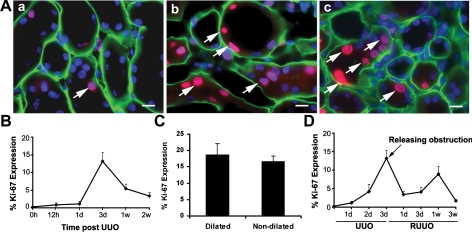
Fig. 1.
Cell proliferation in tubular epithelial cells after unilateral ureteral obstruction (UUO) and reversal of obstruction (RUUO). A: Ki-67 expression (red, arrows) in renal tubular epithelial cells in mice that underwent sham operation (a), 3 days after UUO (b), and 1 wk after RUUO (c). Tubular basement is stained with entactin (green). Nuclei are counterstained with DAPI (blue). Scale bar = 10 μM. B: quantification of Ki-67 expression in tubular epithelial cells after UUO; n = 6. C: there was no significant difference in Ki-67 expression between dilated and nondilated proximal tubules 3 days after UUO (P = 0.3, n = 4). D: quantification of Ki-67 expression in tubular epithelial cells 3 days after UUO followed by RUUO; n = 5. Values are means ± SE.

