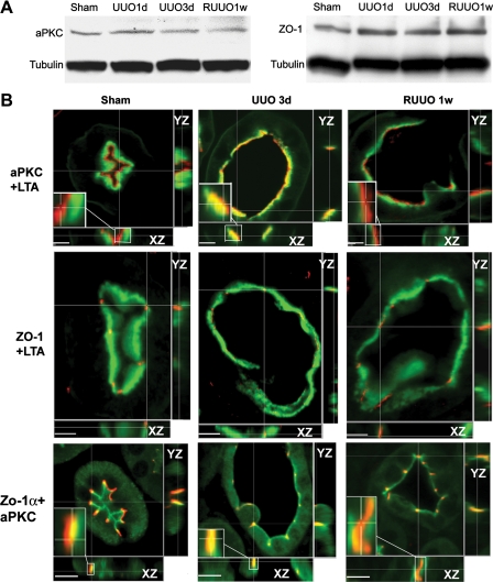
Fig. 4.
Expression and cellular localization of polarity proteins. A: immunoblot blot analysis of atypical PKC (aPKC) and zonula occludens (ZO-1) shows no changes in protein levels in the kidneys after UUO and RUUO. B: laser-scanning confocal microscopic examination of the localization of aPKC and ZO-1 in control proximal tubules, dilated proximal tubules 3 days after UUO, and recovering proximal tubules 1 wk after RUUO. Proximal tubules are stained with Lotus tetragonolobus agglutinin (LTA; green) to reveal the brush border. aPKC (red) is localized to the most apical aspect in the control tubules and the recovering tubules 1 wk after RUUO but merges into the brush-border zone in dilated tubules 3 days after UUO (top). ZO-1 (red) is localized at the apical surface in control tubules and tubules after UUO or RUUO (middle). Costaining of aPKC (green) and ZO-1α+ (red) indicates their colocalization to the tight junctions in the normal and RUUO kidneys. However, aPKC is more subapically localized by further merging into the ZO-1α+ domain in UUO 3-day tubules (bottom). Insets: higher magnification of the localization of aPKC with LTA or ZO-1α+ at the XZ-plane. Scale bars = 5 μm.

