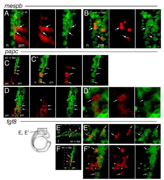
Fig. 3.
Expression of the rostral segment polarity marker genes mespb, papc, and fgf8 by wild-type cells in fused somites/tbx24 hosts. (A–B) Expression of mespb mRNA (red) in confocal sections of the right-hand side of the PM at the A/P level of the segment polarity zone in 8-somite stage embryos containing transplanted wild-type cells (green). (A) Normal mespb expression in wild-type embryos, arrows indicate transplanted cells expressing mespb. (B) Cell-autonomous mespb expression in wild-type donor cells in fss/tbx24 host PSM (arrows). (C–D’) Expression of papc mRNA (red) in confocal sections of the paraxial mesoderm at an A/P level spanning the segment polarity zone in 8-somite stage fss/tbx24 mutant host embryos containing transplanted wild-type cells (green). (C) papc expression associated with small wild-type donor cell clusters (arrows), in contrast to absence of papc on contralateral side. (C’) Higher magnification of C showing papc expression only in wild-type donor cells (arrows). (D) Striped expression of papc in large, high-density clone of wild-type cells (arrows) and location of papc expression in host cell (arrowhead). (D’) Higher magnification of region indicated by arrowhead in D showing papc expression in fss/tbx24 host cell (arrow). (E–F’) Expression of fgf8 mRNA (red) in confocal sections of the paraxial mesoderm at an A/P level spanning the segment polarity zone in an 18-somite (lateral view E, E’) and 6-somite stage (dorsal view F, F’) fss/tbx24 mutant host embryos containing transplanted wild-type cells (green). Location of E, E’ shown in diagrammatic form. (E) fgf8 expression in wild-type cells (arrows), dashed line indicates the dorsal extent of the embryo, the dotted lines delimit the paraxial mesoderm, and the asterisk marks the intermediate mesoderm. (E’) Higher magnification of fgf8 expressing region in panel C, arrows mark fgf8-positive wild-type donor cells. (F) Expression of fgf8 associated with wild-type donor cells in paraxial mesoderm, notochord delineated with dashed line and position of the lateral edge of embryo with a dotted line. (F’) Higher magnification of F, showing fgf8 expression in wild-type donor (arrows) and fss/tbx24 mutant host cells (arrowheads). c = central nervous system, m = paraxial mesoderm, y = yolk, n = notochord, pm = paraxial mesoderm.

