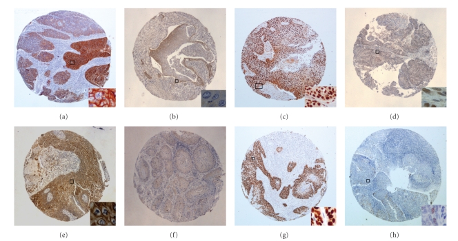
Figure 2.
Immunohistochemistry performed on tissue microarrays. (a) EGFR protein expression in all tumor cells with focal intense complete membranous staining (+3); (b) EGFR negative case showing mild cytoplasmic focal staining; (c) ERCC1 protein strong nuclear positivity; (d) ERCC1 protein expression with equal intensity in neoplastic cells and stromal fibroblasts (regarded as negative staining); (e) MET strong cytoplasmic and membraneous protein expression; (f) Lack of MET protein expression in tumor cells; (g) p-53 strong nuclear protein expression; (h) p-53 expression in a small fraction of tumor cells (regarded as negative staining). Original magnification x20; insets (a), (c), and (g) x200; insets (b), (d), (e), and (h) x400.

