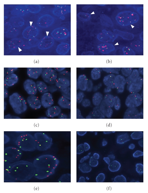
Figure 3.
Fluorescence in situ hybridization with gene and centromeric specific probes. (a) and (b) Neoplastic nuclei showing polysomy of chromosome 7 (CEP7, green signals) and EGFR high level gene gain (red signals, arrowheads); (c) Neoplastic nuclei showing trisomy or polysomy of the MET gene (red signals) and SE7 (green signals); (d) Representative area from a case without genetic alterations. The majority of the neoplastic nuclei have 2 copies of the MET gene and SE7; (e) High polysomy of the D7S486 locus (red signals); (f) Deletion of the D7S486 gene locus in tumor cells, as defined by the presence of a single gene locus probe signal (red signals) and two CEP7 signals (green signals), or by the simultaneous lack of both of the gene locus signals and the presence of CEP7 signals (hemizygous and homozygous deletion, resp.).

