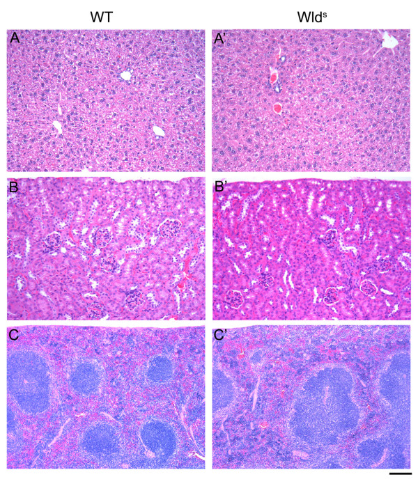
Figure 4.
WldS protein expression did not affect histopathology of non-neuronal organs. H & E staining of tissues showed no obvious qualitative differences between wild type (left), heterozygous (data not shown) or homozygous WldS mice (right) for liver (A), kidney (B), and spleen (C). The selected organs shown represent non-neuronal organs with low (liver), medium (kidney) and high (spleen) levels of WldS protein expression. Scale bar = 100 μm (A&B), 200 μm (C).

